Medial Ulnar Collateral Ligament Injury at the Elbow
Contents
- 1 What is the Ulnar Collateral Ligament?
- 2 What causes Ulnar Collateral Ligament Injury?
- 3 What are the symptoms of Ulnar Collateral Ligament Injury?
- 4 How is Ulnar Collateral Ligament Injury diagnosed?
- 5 How is Ulnar Collateral Ligament Injury treated?
- 6 Why see Dr. Knight for medial collateral ligament surgery?
- 7 Animated Videos
What is the Ulnar Collateral Ligament?
The elbow is a complicated hinge style joint. The humerus (long bone in the upper arm) must articulate with the radius and ulna (two bones in the forearm). There are several ligaments that unite to form a joint capsule linking these bones at the elbow. The Ulnar Collateral Ligament (UCL) is located on the inner aspect of the elbow connecting the humerus to the ulna. Together, the UCL and Lateral Collateral Ligament (LCL) are the primary source of joint stability at the elbow. The UCL is also known as the Medial Collateral Ligament.
What causes Ulnar Collateral Ligament Injury?
Most commonly, the UCL is injured gradually from recurrent stressors on the ligament. Athletes that perform repetitive throwing or overhead motions are the most at risk. Over time microtears may develop leading to eventual rupture. Trauma or dislocation of the elbow may also stretch, fray or tear the ligament.
What are the symptoms of Ulnar Collateral Ligament Injury?
In an acute rupture the athlete may experience a popping sensation followed by a sharp pain to the inside aspect of the elbow after a particularly forceful throw and the subsequent inability to continue throwing. In chronic UCL injury the pain may present after a session of overhead or throwing activity with decreasing ability to perform the movement at full strength. There may be bruising or swelling on the inside of the elbow and clenching the fist distal to injury may be painful. Some patients report tingling or numbness to the ring and/or pinky finger if the ulnar nerve is irritated. UCL injury rarely affects the ability to perform everyday tasks such as carrying groceries or lifting small objects.
How is Ulnar Collateral Ligament Injury diagnosed?
Diagnosis is often based on patient history and symptoms. A physical examination will reveal laxity in the ligament, areas of tightness, crepitation (cracking or popping), or pain. A Valgus Stress Test evaluates the stability of the affected elbow. Various imaging studies may be ordered to assist in diagnosis.
X-rays may be taken to assess for fracture, bone spurs or calcification of the ligament. Valgus Stress radiographs may be taken if the joint is partially dislocated. Ultrasounds, computed tomography (CT) scans and/or magnetic resonance imaging (MRI) using contrast dye may be used to visualize the ligament and diagnose rupture. Since these tests aren’t always able to show a problem if there is one, some surgeons will perform arthroscopy (tiny camera inserted into joint through a small incision) to visualize the injury for a definitive diagnosis.
How is Ulnar Collateral Ligament Injury treated?
Non-surgical
Treatment depends on the severity of injury and symptoms. Elbow instability can be successfully treated without invasive surgery. Activity modification and/or rest will be required to allow the damage to heal. Ice may be applied periodically in the initial phase of treatment. Anti-inflammatory and analgesic medications may be used to combat inflammation and pain. Once the inflammation has subsided, therapy will be used to strengthen the surrounding muscles for joint stability.
Surgical
If conservative treatment fails, surgical intervention may be required. The surgeon will either repair or replace (reconstruct) the ligament. Repairs are performed in the case of avulsion, where the ligament is pulled away from its attachment to the humerus. This is rarely the case. More frequently a ligament reconstruction surgery is performed called Tommy John surgery after the first athlete to receive the procedure in 1974. In Tommy John Surgery a donor tendon is harvested and used to re-anchor the bones together at the original sites of UCL attachment using proven techniques.
Post-operatively the elbow is immobilized in a stiff brace for 1 – 2 weeks. A hinged brace follows along with limited exercise and therapy. Full recovery can be anticipated at approximately 12 – 18 months after surgery.
Why see Dr. Knight for medial collateral ligament surgery?
Dr. John Knight is a board certified, Fellowship-trained orthopedic surgeon with more than 25 years of experience in orthopedic surgery. Dr. Knight is well-versed in the latest orthopedic surgical techniques, which feature minimally invasive surgery with the best recovery of function over time. A medial ulnar collateral ligament injury can severely affect your ability to throw, which could impact your livelihood or your enjoyment of amateur sports. Choosing the right surgeon is an essential part of your treatment.
Dr. Knight is one of the premier hand surgeons in Dallas. We invite you to visit him at our Southlake office or Dallas office today.
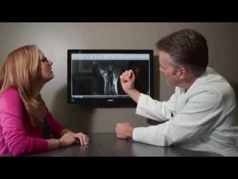
Animated Videos
Disclaimer
HandAndWristInstitute.com does not offer medical advice. The information presented here is offered for informational purposes only. Read Disclaimer
Dr. John Knight
Dr. Knight is a renowned hand, wrist and upper extremity surgeon with over 25 years of experience. Dr. Knight is a Board Certified Orthopedic Surgeon and Fellowship trained. Dr Knight has appeared on CNN, The Doctors TV, Good Morning America, The Wall Street Journal, The Washington Post, Forbes, The Huffington Post, Entrepreneur, Oxygen network and more.
Schedule Appointment

Meet the Doctor

Where Does it Hurt?®
Not sure what service you need or what injury or syndrome you may have? Use our free, interactive tool to help you understand more about what you are experiencing. Start by clicking on the image below.
Real Patient Reviews

“I don’t often write reviews for Doctors offices..But this office is really exceptional in terms of service and my wrist is now great! Which is really the most important thing.”
Featured On