How Hard Do You Have to Hit Your Elbow to Fracture It?
What is an elbow fracture?
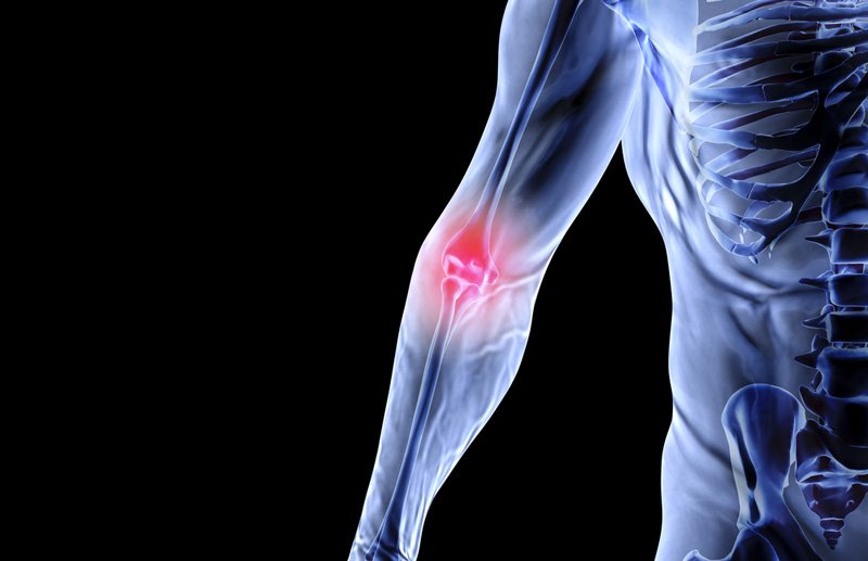
The elbow is made up of three types of bones: the humerus (bone of the upper arm), the radius and ulna (bones of the lower arm). Each bone contributes to what is considered the “elbow”, however, the olecranon process of the ulna is the prominent portion of the elbow. The olecranon protrusion is the bony point felt when the elbow is touched.
Elbow fracture also called olecranon fracture are common (comprises approximately 10% of all fractures around the elbow), and it involves trauma to the bony point of the elbow (called the olecranon process). Elbow fractures vary in complexity from simple transverse fractures to oblique fractures, and comminuted fractures.
What are the causes of an elbow fracture?
Direct trauma such as a hard hit, for example with a baseball bat, at the elbow, is a common cause of elbow fracture among young children and adults. Also, falling directly on the elbow can result in a comminuted fracture, or the bones of the elbow breaking into tiny fragments. Indirect trauma such as falling with an outstretched arm is often the cause of elbow fracture among the elderly.
What are the symptoms of an elbow fracture?
The symptoms of an elbow fracture include intense pain, which is often localized to the posterior elbow. Swelling following a fracture limits people from moving their elbow and can cause a reduction in the range of motion (ROM) at the elbow. Bruising around the elbow can occur – bruising can travel along the arm towards the hand or shoulder. Symptoms can also include numbness in the fingers and tenderness to touch.
How is an elbow fracture diagnosed?
Diagnosing an elbow fracture involves obtaining a detailed medical history. Doctors will perform a physical examination of the elbow and arm. Doctors will check for skin cuts and lacerations. In more severe fractures, bone fragments can cut through the skin at the elbow.
Other examinations may include evaluating the fingers and arms for sensation. Physicians will assess other parts of the arm to rule out any other damage.
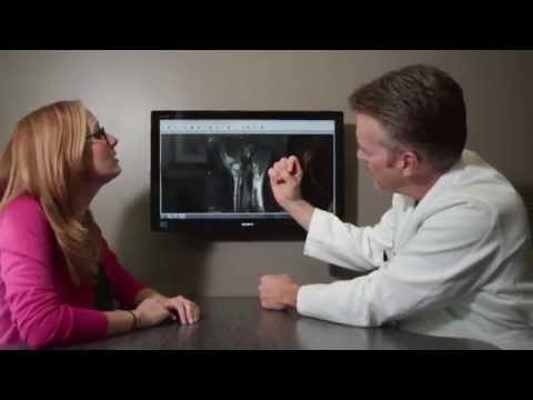
X-ray images or radiographs can provide a better picture of underlying trauma to the bones of the elbow. Physicians will request X-rays of the elbow and arm to make a better diagnosis – and rule out other injuries. Radiographs are also important for determining fracture patterns to help with surgical procedures.
Computer tomography (CT) scans can also be combined with X-rays to aid in preoperative planning.
Treatment options for an elbow fracture
There are non-surgical treatment and surgical treatment for an elbow fracture. The treatment for elbow fracture depends on the severity of the fracture.
Nonsurgical Treatment
Doctors will recommend non-steroidal anti-inflammatory drugs (NSAIDs) like ibuprofen to relieve pain symptoms, and also advise applying ice to reduce the swelling. For non-displaced fractures, non-surgical treatment often involves wearing a cast (or splits) to immobilize the elbow.
Splints are usually worn for three (3) to six (6) weeks before they are removed. The splints/casts are designed such that the elbow is immobilized at 45° – 90°. Exercises that improve range of motion are considered to aid movement at the elbow.
Surgical treatment
There are different surgical procedures available for repairing elbow fractures. These procedures depend on the age of the patient, the patient’s pre-existing conditions e.g an elderly patient with osteoporosis, etc. and the severity of the fracture.
Surgery is usually considered for displaced and open elbow fractures. In displaced fractures, the bones of the elbow are have shifted in position during the trauma that hits the elbow. Displaced fractures are often presented by older patients. Open fractures, on the other hand, occurs when the bones of the elbow are broken into two or more tiny pieces.
Because open fractures cut through the skin and expose the patients to infections, surgery must be performed as soon as possible. Patients with open fractures of the elbow are administered antibiotics (intravenously). During surgery, bone fragments are cleaned out and repaired.
Common surgical techniques for repairing elbow fractures are
Tension band fixation or internal fixation
This procedure is commonly used for repairing non-comminuted transverse elbow fractures. In this procedure, the pieces of bones are held together with screws, wires, pins, or metal plates called tension band.
Intramedullary fixation
This technique is similar to the tension band fixation. Used for repairing simple transverse and oblique fractures.
Plate and screw fixation
This procedure is used for repairing comminuted elbow fractures, Monteggia fractures, fracture-dislocations, and oblique fractures. This procedure provides more stability to the bones than the tension band.
Excision and triceps advancement
This surgical treatment is recommended for older patients with osteoporotic bone. It is also performed for fractures with extensive comminution (less than 50% of the joint surface) or non-union fractions. In this technique, the triceps tendon is reattached with nonabsorbable sutures passed through drill holes in the ulna.
Bone graft
Bone grafts are used when bone fragments are not fully recovered. A bone graft can be obtained from the patient e.g. from the hip or a donor. Artificial biocompatible materials can also be used to repair the fracture
Post-operative recovery and management.
The patient must continue to follow the physician’s recommendations after an elbow fracture treatment. Physical therapy and rehabilitation are advised to facilitate recovery and improve range of motion, and hand use.
Exercise therapists can help patients strengthen the muscles of the elbow and forearm, and also decrease stiffness at the joint. Patients should avoid lifting, pushing or pulling heavy objects with the affected arm (if surgery is performed) until the arm has recovered well (usually about six weeks after surgery).
Citations
- S.D.S. Newman, C. Mauffrey, S. Krikler. Olecranon fractures. Injury. Volume 40, Issue 6. 2009. Pages 575-581. ISSN 0020-1383. https://doi.org/10.1016/j.injury.2008.12.013.
- “Elbow (Olecranon) FracturesOrthoInfoAAOS.” orthoinfo.aaos.org/en/diseases–conditions/elbow-olecranon-fractures/.
- “Olecranon Fractures.” 5 Oct. 2016, www.orthobullets.com/trauma/1022/olecranon-fractures.
Read more about Elbow Fractures – https://www.handandwristinstitute.com/elbow-fractures/

Dr. John Knight
Dr. Knight is a renowned hand, wrist and upper extremity surgeon with over 25 years of experience. Dr. Knight is a Board Certified Orthopedic Surgeon and Fellowship trained. Dr Knight has appeared on CNN, The Doctors TV, Good Morning America, The Wall Street Journal, The Washington Post, Forbes, The Huffington Post, Entrepreneur, Oxygen network and more.
Meet the Doctor

Schedule Appointment
New patients can schedule an appointment online and fill out your patient information to save time. Existing patients, click here.

Where Does it Hurt?®
Not sure what service you need or what injury or syndrome you may have? Use our free, interactive tool to help you understand more about what you are experiencing. Start by clicking on the image below.
Virtual Consultation
Located out of the area? Dr. Knight may be able to help you virtually with an online virtual consultation.

Real Patient Reviews

“I don’t often write reviews for Doctors offices..But this office is really exceptional in terms of service and my wrist is now great! Which is really the most important thing.”
Featured On